

Journal of Clinical Pediatrics ›› 2024, Vol. 42 ›› Issue (6): 526-532.doi: 10.12372/jcp.2024.23e0289
• Original Article • Previous Articles Next Articles
LIANG Jiawei, MA Xiaohui, JIA Xuan( )
)
Received:2023-04-11
Published:2024-06-15
Online:2024-06-07
LIANG Jiawei, MA Xiaohui, JIA Xuan. Study on the application value of the new Lake Louise criteria in children with myocarditis[J].Journal of Clinical Pediatrics, 2024, 42(6): 526-532.
"
| 组 别 | 例数 | 年龄[M(P25~P75)]/岁 | 男性[n(%)] | HCT(x±s)/% | 心率(x±s)/次·min-1 | ER(x±s) |
|---|---|---|---|---|---|---|
| 心肌炎组 | 29 | 8.8(4.2~13.4) | 16(55.2) | 37.4±5.1 | 84.6±30.9 | 2.0±0.5 |
| 非心肌炎组 | 50 | 9.7(6.2~13.2) | 27(54.0) | 37.9±3.1 | 79.7±16.8 | 1.6±0.2 |
| 统计量 | Z=0.90 | χ2=0.01 | t=0.49 | t=0.79 | t=4.20 | |
| P | 0.373 | 0.920 | 0.626 | 0.435 | <0.001 |
"
| 组 别 | 例数 | T2 mapping值/ms | T1 native值/ms | T1 enhanced值/ms | ECV/% |
|---|---|---|---|---|---|
| 心肌炎组 | 29 | 60.0±9.5 | 1 185.7±102.7 | 469.3±80.2 | 36.5±7.1 |
| 非心肌炎组 | 50 | 49.5±5.5 | 1 027.4±28.8 | 530.9±64.7 | 26.4±3.3 |
| t值 | 5.43 | 8.12 | 3.73 | 7.21 | |
| P | <0.001 | <0.001 | <0.001 | <0.001 |
"
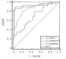
| 项 目 | AUC(95%CI) | P | 截断值 | 灵敏度/% | 特异度/% | 约登指数 |
|---|---|---|---|---|---|---|
| LLC | 0.75(0.64~0.84) | <0.001 | - | 51.7 | 98.0 | 0.50 |
| T2 mapping | 0.86(0.77~0.93) | <0.001 | 52.5 ms | 79.3 | 80.0 | 0.59 |
| T1 native | 0.92(0.84~0.97) | <0.001 | 1 086.0 ms | 82.8 | 100.0 | 0.83 |
| T1 enhanced | 0.70(0.59~0.80) | <0.001 | 516.5 ms | 79.3 | 54.0 | 0.33 |
| ECV | 0.93(0.85~0.98) | <0.001 | 30.4% | 89.7 | 96.0 | 0.86 |
"

"
| 项 目 | 准确度/% | 灵敏度/% | 特异度/% | 阳性预测值/% | 阴性预测值/% |
|---|---|---|---|---|---|
| LLC | 81.0 | 51.7 | 98.0 | 93.8 | 77.8 |
| 新LLC | 92.4 | 89.7 | 94.0 | 89.7 | 94.0 |
| χ2值 | 4.45 | 10.07 | 1.04 | 0.00 | 5.74 |
| P | 0.035 | 0.002 | 0.307 | 1.000 | 0.017 |
| [1] | 陈燕, 罗琳, 何健龙, 等. MRI T1 mapping、T2 mapping心肌分段在诊断急性心肌炎中的应用[J]. 中国医学影像学杂志, 2019, 27(8): 599-604. |
| [2] | Haaf P, Garg P, Messroghli DR, et al. Cardiac T1 Mapping and Extracellular Volume (ECV) in clinical practice: a comprehensive review[J]. J Cardiovasc Magn Reson, 2016, 18(1): 89. |
| [3] |
Friedrich MG, Sechtem U, Schulz-Menger J, et al. Cardiovascular magnetic resonance in myocarditis: a JACC white paper[J]. J Am Coll Cardiol, 2009, 53(17): 1475-1487.
doi: 10.1016/j.jacc.2009.02.007 pmid: 19389557 |
| [4] | Schumm J, Greulich S, Wagner A, et al. Cardiovascular magnetic resonance risk stratification in patients with clinically suspected myocarditis[J]. J Cardiovasc Magn Reson, 2014, 16(1): 14. |
| [5] | 周笛, 庄白燕, 赵世华, 等. 心血管MR诊断心肌炎研究进展: 基于2018《非缺血性心肌炎症诊断CMR标准修改》专家推荐意见[J]. 中国医学影像技术, 2019, 35(10): 1574-1577. |
| [6] |
Radunski UK, Lund GK, Stehning C, et al. CMR in patients with severe myocarditis: diagnostic value of quantitative tissue markers including extracellular volume imaging[J]. JACC Cardiovasc Imaging, 2014, 7(7): 667-675.
doi: 10.1016/j.jcmg.2014.02.005 pmid: 24954462 |
| [7] | Bohnen S, Radunski UK, Lund GK, et al. Performance of t1 and t2 mapping cardiovascular magnetic resonance to detect active myocarditis in patients with recent-onset heart failure[J]. Circ Cardiovasc Imaging, 2015, 8(6): e003073. |
| [8] | Liu G, Yang X, Su Y, et al. Cardiovascular magnetic resonance imaging findings in children with myocarditis[J]. Chin Med J (Engl), 2014, 127(21): 3700-3705. |
| [9] |
Dass S, Suttie JJ, Piechnik SK, et al. Myocardial tissue characterization using magnetic resonance noncontrast t1 mapping in hypertrophic and dilated cardiomyopathy[J]. Circ Cardiovasc Imaging, 2012, 5(6): 726-733.
doi: 10.1161/CIRCIMAGING.112.976738 pmid: 23071146 |
| [10] | 赵世华. 迎接心脏磁共振成像新技术挑战[J]. 中国医学影像技术, 2017, 33(8): 1125-1128. |
| [11] |
Luetkens JA, Homsi R, Sprinkart AM, et al. Incremental value of quantitative CMR including parametric mapping for the diagnosis of acute myocarditis[J]. Eur Heart J Cardiovasc Imaging, 2016, 17(2): 154-161.
doi: 10.1093/ehjci/jev246 pmid: 26476398 |
| [12] |
Ferreira VM, Schulz-Menger J, Holmvang G, et al. Cardiovascular magnetic resonance in nonischemic myocardial inflammation: expert recommendations[J]. J Am Coll Cardiol, 2018, 72(24): 3158-3176.
doi: S0735-1097(18)38843-0 pmid: 30545455 |
| [13] | 中华医学会儿科学分会心血管学组, 中华医学会儿科学分会心血管学组心肌炎协作组, 中华儿科杂志编辑委员会, 等. 儿童心肌炎诊断建议(2018年版)[J]. 中华儿科杂志, 2019, 57(2): 87-89. |
| [14] | Youn JC, Hong YH, Lee HJ, et al. Contrast-enhanced T1 mapping-based extracellular volume fraction independently predicts clinical outcome in patients with non-ischemic dilated cardiomyopathy: a prospective cohort study[J]. Eur Radiol, 2017, 27(9): 3924-3933. |
| [15] | 刘钢, 张兴梅, 温兆赢, 等. 心脏磁共振定量组织标记技术对急性心肌炎的诊断价值[J]. 中国医药, 2017, 12(1): 41-45. |
| [16] | 陈文剑, 郭婷婷, 张继良, 等. 心脏磁共振对心肌炎的诊断价值[J]. 中国实用医刊, 2020, 47(9): 5-7. |
| [17] | 欧阳海春, 陈海雄, 胡允兆, 等. 磁共振在急性病毒性心肌炎中的诊断价值[J]. 中华心血管病杂志, 2014, 42(11): 927-931. |
| [18] | Dabir D, Vollbrecht TM, Luetkens JA, et al. Multiparametric cardiovascular magnetic resonance imaging in acute myocarditis: a comparison of different measurement approaches[J]. J Cardiovasc Magn Reson, 2019, 21(1): 54. |
| [19] |
Bami K, Haddad T, Dick A, et al. Noninvasive imaging in acute myocarditis[J]. Curr Opin Cardiol, 2016, 31(2): 217-223.
doi: 10.1097/HCO.0000000000000265 pmid: 26731291 |
| [20] | 万俊义. 心脏磁共振钆对比剂延迟强化的临床意义及判断预后的价值[J]. 中国医学影像技术, 2012, 28(8): 1600-1603. |
| [21] | Banka P, Robinson JD, Uppu SC, et al. Cardiovascular magnetic resonance techniques and findings in children with myocarditis: a multicenter retrospective study[J]. J Cardiovasc Magn Reson, 2015, 17: 96. |
| [22] |
Lurz P, Eitel I, Adam J, et al. Diagnostic performance of CMR imaging compared with EMB in patients with suspected myocarditis[J]. JACC Cardiovasc Imaging, 2012, 5(5): 513-524.
doi: 10.1016/j.jcmg.2011.11.022 pmid: 22595159 |
| [23] |
Mavrogeni S, Bratis K, Georgakopoulos D, et al. Evaluation of myocarditis in a pediatric population using cardiovascular magnetic resonance and endomyocardial biopsy[J]. Int J Cardiol, 2012, 160(3): 192-195.
doi: 10.1016/j.ijcard.2011.04.019 pmid: 21561672 |
| [24] |
Ferreira VM, Piechnik SK, Dall’Armellina E, et al. T(1) mapping for the diagnosis of acute myocarditis using CMR: comparison to T2-weighted and late gadolinium enhanced imaging[J]. JACC Cardiovasc Imaging, 2013, 6(10): 1048-1058.
doi: S1936-878X(13)00539-1 pmid: 24011774 |
| [25] | Thavendiranathan P, Walls M, Giri S, et al. Improved detection of myocardial involvement in acute inflammatory cardiomyopathies using T2 mapping[J]. Cire Cardiovasc Imaging, 2012, 5(1): 102-110. |
| [26] | 殷亮, 喻思思, 龚良庚. 心脏磁共振纵向弛豫时间定量成像技术进展[J]. 中华放射学杂志, 2016, 50(1): 76-78. |
| [27] | Park CH, Choi EY, Kwon HM, et al. Quantitative T2 mapping for detecting myocardial edema after reperfusion of myocardial infarction: validation and comparison with T2-weighted images[J]. Int J Cardiovasc Imaging, 2013, 29(suppl 1): 65-72. |
| [28] |
Ding H, Fernandez-de-Manuel L, Schär M, et al. Three-dimensional whole-heart T2 mapping at 3T[J]. Magn Reson Med, 2015, 74(3): 803-816.
doi: 10.1002/mrm.25458 pmid: 25242141 |
| [29] | Lee JJ, Liu S, Nacif MS, et al. Myocardial T1 and extracellular volume fraction mapping at 3 tesla[J]. J cardiovasc Magn Reson, 2011, 13(1): 75. |
| [30] | Kawel N, Nacif M, Zavodni A, et al. T1 mapping of the myocardium: intra-individual assessment of the effect of field strength, cardiac cycle and variation by myocardial region[J]. J Cardiovasc Magn Reson, 2012, 14(1): 27. |
| [31] | Nadjiri J, Nieberler H, Hendrich E, et al. Performance of native and contrast- enhanced T1 mapping to detect myocardial damage in patients with suspected myocarditis: a head-to-head comparison of different cardiovascular magnetic resonance techniques[J]. Int J Cardiovasc Imaging, 2017, 33(4): 539- 547. |
| [32] | Wassmuth R, Prothmann M, Utz W, et al. Variability and homogeneity of cardiovascular magnetic resonance myocardial T2-mapping in volunteers compared to patients with edema[J]. J Cardiovasc Magn Reson, 2013, 15(1): 27. |
| [33] | 刘新峰, 马海彦, 王荣品, 等. T1 mapping和细胞外容积分数技术在急性病毒性心肌炎的临床应用[J]. 实用医学杂志, 2019, 35(5): 800-803. |
| [34] | Dabir D, Child N, Kalra A, et al. Reference values for healthy human myocardium using a T1 mapping methodology: results from the international T1 multicenter cardiovascular magnetic resonance study[J]. J Cardiovasc Magn Reson, 2014, 16(1): 69. |
| [35] |
Miller CA, Naish JH, Bishop P, et al. Comprehensive validation of cardiovascular magnetic resonance techniques for the assessment of myocardial extracellular volume[J]. Circ Cardiovasc Imaging, 2013, 6(3): 373-383.
doi: 10.1161/CIRCIMAGING.112.000192 pmid: 23553570 |
| [36] | Cornicelli MD, Rigsby CK, Rychlik K, et al. Diagnostic performance of cardiovascular magnetic resonance native T1 and T2 mapping in pediatric patients with acute myocarditis[J]. J Cardiovasc Magn Reson, 2019, 21(1): 40. |
| [37] |
Lurz P, Luecke C, Eitel I, et al. Comprehensive cardiac magnetic resonance imaging in patients with suspected myocarditis: the MyoRacer-trial[J]. J Am Coll Cardiol, 2016, 67(15): 1800-1811.
doi: S0735-1097(16)00610-0 pmid: 27081020 |
| [38] | Baeßler B, Schaarschmidt F, Dick A, et al. Mapping tissue inhomogeneity in acute myocarditis: a novel analytical approach to quantitative myocardial edema imaging by T2-mapping[J]. J Cardiovasc Magn Reson, 2015, 17: 115. |
| [39] | Luetkens JA, Homsi R, Dabir D, et al. Comprehensive cardiac magnetic resonance for short-term follow-up in acute myocarditis[J]. J Am Heart Assoc, 2016, 5(7): e003603. |
| [1] | LUO Mingjing, YU Jiaming, WANG Xiaodong, ZHANG Xiaoling, YU Yue, ZHANG Yu, WEN Feiqiu, LIU Sixi. Clinical analysis of invasive fungal disease secondary to allogeneic hematopoietic stem cell transplantation in 424 children with thalassemia [J]. Journal of Clinical Pediatrics, 2025, 43(1): 21-28. |
| [2] | LIU Dongxia, JIN Rong, LIN Rongjun. Risk factors analysis of severe refractory Mycoplasma pneumoniae pneumonia complicated with bronchitis obliterans in children [J]. Journal of Clinical Pediatrics, 2025, 43(1): 29-34. |
| [3] | ZHONG Jinhong, WANG Can, CHEN Fang. Progress in the research of infantile fiberoptic bronchoscopy sedation [J]. Journal of Clinical Pediatrics, 2025, 43(1): 50-55. |
| [4] | JIANG Weiqin, WANG Jing, CHENG Anna, CHEN Tingting, HUANG Yujuan. Predictors of recurrent febrile seizures during the same febrile illness in children with febrile seizures [J]. Journal of Clinical Pediatrics, 2025, 43(1): 8-13. |
| [5] | QIU Xiu, WEI Dongmei, LIN Shanshan, XIA Huimin, ZHOU Wenhao. Principles and practice of the Born in Guangzhou Cohort Study [J]. Journal of Clinical Pediatrics, 2024, 42(9): 747-752. |
| [6] | FAN Jianxia. The origins and development of the healthy life trajectory program: a cohort of community-family-mother-child multidimensional interventions for overweight and obesity in children [J]. Journal of Clinical Pediatrics, 2024, 42(9): 768-773. |
| [7] | JIANG Tao, LI Shuangjie, TANG Lian, OUYANG Wenxian. Immunobiological properties of peripheral blood MAIT cells in children with chronic hepatitis B [J]. Journal of Clinical Pediatrics, 2024, 42(9): 787-790. |
| [8] | ZHOU Jie, LIU Keqiang, WANG Jinling, WANG Ying. Megacystis-microcolon-intestinal hypoperistalsis syndrome caused by MYH11 elongating mutation : a case report and literatures review [J]. Journal of Clinical Pediatrics, 2024, 42(9): 798-804. |
| [9] | CHU Sijia, TANG Jihong. Research progress of central nervous system injury associated with pediatric acute lymphoblastic leukemia and its treatment [J]. Journal of Clinical Pediatrics, 2024, 42(9): 811-816. |
| [10] | DING Yaping, XIA Shanshan, ZHANG Chenmei. Interpretation of “2023 Children’s Renal Nutrition Working Group Clinical Practice Recommendations: Nutritional Management of Children with Acute Kidney Injury” [J]. Journal of Clinical Pediatrics, 2024, 42(8): 667-672. |
| [11] | LI Yirong, LI Huiping, GAO Jingyu, XIAO Yuhua, CHEN Xiaomin, LU Yanling, ZHAO Nana, FENG Xiaoqin. Comparison of different doses of cytarabine for induction chemotherapy in children with acute myeloid leukemia in FLAG-IDA regimen [J]. Journal of Clinical Pediatrics, 2024, 42(8): 673-677. |
| [12] | HUANG Bo, DONG Yanying, SONG Linlan. Clinical characteristics of 348 children with infectious mononucleosis [J]. Journal of Clinical Pediatrics, 2024, 42(8): 678-683. |
| [13] | WANG Dan, SHAO Jingbo, LI Hong, ZHANG Na, ZHU Jiashi, FU Pan, WANG Zhen. Clinical analysis of 38 cases of hematological malignancies complicated with tumor lysis syndrome in children [J]. Journal of Clinical Pediatrics, 2024, 42(8): 684-690. |
| [14] | MA Yan, WEI Xingjiao, BAI Hua, ZHANG Yan, TIAN Xinmin, Aqsa Ahmad, LIANG Lijun. Analysis of etiological composition and clinical features of stage 5 chronic kidney disease in children in a tertiary hospital in western China [J]. Journal of Clinical Pediatrics, 2024, 42(8): 697-703. |
| [15] | WANG Ye, ZHANG Linlin, CHI Zuofei, SUN Ruowen, JIANG Zehui, XU Gang. A case of clinical report of T-lymphoblastic lymphoma secondary to acute promyelocytic leukemia in children [J]. Journal of Clinical Pediatrics, 2024, 42(8): 722-727. |
|
||