

Journal of Clinical Pediatrics ›› 2023, Vol. 41 ›› Issue (9): 674-679.doi: 10.12372/jcp.2023.22e0450
• Nervous System Disease • Previous Articles Next Articles
HOU Ruolin, WU Jing( ), LI Ling()
Received:2022-04-18
Published:2023-09-15
Online:2023-09-05
Contact:
WU Jing,LI Ling
E-mail:wujing01@xinhuamed.com.cn;liling@xinhuamed.com.cn
HOU Ruolin, WU Jing, LI Ling. Pediatric autoimmune encephalitis with brain MRI showing meningeal thickening and enhancement[J].Journal of Clinical Pediatrics, 2023, 41(9): 674-679.
"
"
| 项 目 | 病例1 | 病例2 | |
|---|---|---|---|
| 第一次入院 | 第二次入院 | ||
| 血自身抗体系列 | dsDNA、抗SSA弱阳性,Ro-52阳性 | ANA核型颗粒型、ANA(1:40)阳性,Ro-52弱阳性 | 阴性 |
| 脑脊液检测 | |||
| 细胞数/mm3 | 6↑ | 5 | 6↑ |
| 体液蛋白定量/mg·L-1 | 691.5↑ | 178.4 | 617.6↑ |
| 葡萄糖/mmol·L-1 | 3.76 | 4.39 | 8.67 |
| 氯化物/mmol·L-1 | 125 | 130 | 127.9 |
| IgG/mg·L-1 | 68.9↑ | 10.7 | 34.1 |
| IgA/mg·L-1 | 5.32↑ | 0.67 | 3.17↑ |
| IgM/mg·L-1 | 3.72↑ | 0.15 | 1.74 |
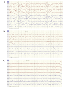
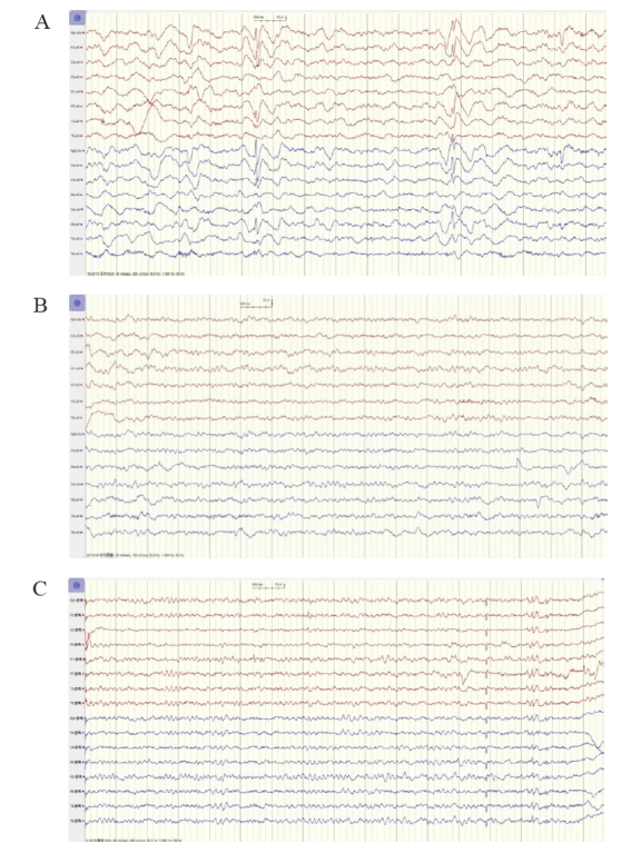
| 脑电图 | 慢波(3.5~4.0 Hz慢波背景,夹杂1.5~2.5 Hz δ波)伴痫样放电 | 背景慢波(6~7 Hz) | 背景慢波(6~8 Hz) |
"
"
| [1] |
Lancaster E, Dalmau J. Neuronal autoantigens: patho-genesis, associated disorders and antibody testing[J]. Nat Rev Neurol, 2012, 8(7): 380-390.
doi: 10.1038/nrneurol.2012.99 pmid: 22710628 |
| [2] |
Li XJ, Hou C, Wu WL, et al. Pediatric anti-N-methyl-D-aspartate receptor encephalitis in southern China: analysis of 111 cases[J]. J Neuroimmunol, 2021, 352: 577479.
doi: 10.1016/j.jneuroim.2021.577479 |
| [3] |
Wang R, Lai XH, Liu X, et al. Brain magnetic resonance-imaging findings of anti-N-methyl-D-aspartate receptor encephalitis: a cohort follow-up study in Chinese patients[J]. Neurol, 2018, 265(2): 362-369.
doi: 10.1007/s00415-017-8707-5 |
| [4] |
Zhang T, Duan Y, Ye J, et al. Brain MRI characteristics of patients with anti-N-methyl-D aspartate receptor encephalitis and their associations with 2-year clinical outcome[J]. AJNR Am J Neuroradiol, 2018, 39(5): 824-829.
doi: 10.3174/ajnr.A5593 |
| [5] |
Xiao X, Fu D, Feng L. Hypertrophic pachymeningitis in a southern Chinese population: a retrospective study[J]. Front Neurol, 2020, 11: 565088.
doi: 10.3389/fneur.2020.565088 |
| [6] |
Cellucci T, Van Mater H, Graus F, et al. Clinical approach to the diagnosis of autoimmune encephalitis in the pediatric patient[J]. Neurol Neuroimmunol Neuroinflamm, 2020, 7(2): e663.
doi: 10.1212/NXI.0000000000000663 |
| [7] |
Antony J, Hacking C, Jeffree RL. Pachymeningeal enhancement-a comprehensive review of literature[J]. Neurosurg Rev, 2015, 38(4): 649-659.
doi: 10.1007/s10143-015-0646-y pmid: 26264063 |
| [8] |
D’Antona L, Jaime Merchan MA, Vassiliou A, et al. Clinical presentation, investigation findings, and treatment outcomes of spontaneous intracranial hypotension syndrome: a systematic review and meta-analysis[J]. JAMA Neurol, 2021, 78(3): 329-337.
doi: 10.1001/jamaneurol.2020.4799 pmid: 33393980 |
| [9] | Tian CL, Pu CQ. Dural enhancement detected by magnetic resonance imaging reflecting the underlying causes of cerebral venous sinus thrombosis[J]. Chin Med J (Engl), 2012, 125(8): 1513-1516. |
| [10] |
Fang B, McKeon A, Hinson SR, et al. Autoimmune glial fibrillary acidic protein astrocytopathy: a novel meningoencephalomyelitis[J]. JAMA Neurol, 2016, 73(11): 1297-1307.
doi: 10.1001/jamaneurol.2016.2549 pmid: 27618707 |
| [11] |
Britton PN, Dale RC, Blyth CC, et al. Causes and clinical features of childhood encephalitis: a multicenter, prospective cohort study[J]. Clin Infect Dis, 2020, 70(12): 2517-2526.
doi: 10.1093/cid/ciz685 pmid: 31549170 |
| [12] |
Bitnun A, Ford-Jones EL, Petric M, et al. Acute childhood encephalitis and Mycoplasma pneumoniae[J]. Clin Infect Dis, 2001, 32(12): 1674-1684.
pmid: 11360206 |
| [13] |
Meyer Sauteur PM, Jacobs BC, Spuesens EB, et al. Antibody responses to Mycoplasma pneumoniae: role in pathogenesis and diagnosis of encephalitis?[J]. PLoS Pathog, 2014, 10(6): e1003983.
doi: 10.1371/journal.ppat.1003983 |
| [14] |
Lin JJ, Hsia SH, Wu CT, et al. Mycoplasma pneumoniae-related postencephalitic epilepsy in children[J]. Epilepsia, 2011, 52(11): 1979-1985.
doi: 10.1111/epi.2011.52.issue-11 |
| [15] |
Lin JJ, Lin KL, Hsia SH, et al. Analysis of status epilepticus with Mycoplasma pneumoniae Encephalitis[J]. Pediatr Neurol, 2010, 43(1): 41-45.
doi: 10.1016/j.pediatrneurol.2010.02.017 |
| [16] |
Narita M. Pathogenesis of neurologic manifestations of Mycoplasma pneumoniae Infection[J]. Pediatr Neurol, 2009, 41(3): 159-166.
doi: 10.1016/j.pediatrneurol.2009.04.012 |
| [17] | Meyer Sauteur PM, Moeller A, Relly C, et al. Swiss national prospective surveillance of paediatric Mycoplasma pneumoniae-associated encephalitis[J]. Swiss Med Wkly, 2016, 146: w14222. |
| [18] |
Li Q, Fu N, Han Y, et al. Pediatric autoimmune encephalitis and its relationship with infection[J]. Pediatr Neurol, 2021, 120: 27-32.
doi: 10.1016/j.pediatrneurol.2021.04.001 pmid: 33964702 |
| [19] |
Venancio P, Brito MJ, Pereira G, et al. Anti-N-methyl-D-aspartate receptor encephalitis with positive serum antithyroid antibodies, IgM antibodies against Mycoplasma pneumoniae and human herpesvirus 7 PCR in the CSF[J]. Pediatr Infect Dis J, 2014, 33(8): 882-883.
doi: 10.1097/INF.0000000000000408 |
| [20] |
Abrantes FF, Moraes MPM, Rezende Filho FM, et al. A clinical approach to hyprtrophic pachymeningitis[J]. Arq Neuropsiquiatr, 2020, 78(12): 797-804.
doi: 10.1590/0004-282x20200073 |
| [21] | Bi Z, Shang K, Cao J, et al. Hypertrophic pachymeningitis in Chinese patients: presentation, radiological findings, and clinical course[J]. Biomed Res Int, 2020, 2020: 2926419. |
| [22] |
Margoni M, Barbareschi M, Rozzanigo U, et al. Idiopathic hypertrophic cranial pachymeningitis as a rare cause of status epilepticus[J]. Neurol Sci. 2019, 40(10): 2193-2195.
doi: 10.1007/s10072-019-03954-9 pmid: 31154557 |
| [23] |
De Virgilio A, de Vincentiis M, Inghilleri M, et al. Idiopathic hypertrophic pachymeningitis: an autoimmune IgG4-related disease[J]. Immunol Res, 2017, 65(1): 386-394.
doi: 10.1007/s12026-016-8863-1 pmid: 27592235 |
| [24] |
Cação G, Calejo M, Alves JE, et al. Clinical features of hypertrophic pachymeningitis in a center survey[J]. Neurol Sci, 2019, 40(3): 543-551.
doi: 10.1007/s10072-018-3689-3 pmid: 30588552 |
| [25] |
Jia H, Xie X, Qi F, et al. Anti-NMDAR encephalitis with simultaneous hypertrophic pachymeningitis in a 68-year-old male: a rare case report[J]. BMC Neurol, 2019, 19(1): 215.
doi: 10.1186/s12883-019-1444-x pmid: 31472692 |
| [26] |
Ueno T, Kon T, Kaneko K, et al. Contrast enhancement of hypertrophic dura mater in MOG antibodyassociated disease[J]. Neurology, 2019, 93(6): 271-272.
doi: 10.1212/WNL.0000000000007909 |
| [27] |
Papathanasiou A, Yeo JM, Humberstone M, et al. MOG antibody-associated hypertrophic pachymeningitis[J]. Mult Scler Relat Disord, 2020, 42: 102074.
doi: 10.1016/j.msard.2020.102074 |
| [28] |
Zhang TY, Cai MT, Zheng Y, et al. Anti-alpha-amino-3-hydroxy-5-methyl-4-isoxazo lepropionic acid receptor encephalitis: a review[J]. Front Immunol, 2021, 12: 652820.
doi: 10.3389/fimmu.2021.652820 |
| [29] |
Diering GH, Huganir RL. The AMPA receptor code of synaptic plasticity[J]. Neuron, 2018, 100(2): 314-329.
doi: S0896-6273(18)30906-1 pmid: 30359599 |
| [30] |
Alves de Lima K, Rustenhoven J, Kipnis J. Meningeal immunity and its function in maintenance of the central nervous system in health and disease[J]. Annu Rev Immunol, 2020, 38: 597-620.
doi: 10.1146/annurev-immunol-102319-103410 pmid: 32340575 |
| [31] |
Papadopoulos Z, Herz J, Kipnis J. Meningeal lymphatics: from anatomy to central nervous system immune surveil-lance[J]. J Immunol. 2020, 204(2): 286-293.
doi: 10.4049/jimmunol.1900838 pmid: 31907271 |
| [32] |
Tan CB, Zhong M, Yao ZX, et al. Anti-GFAP antibody-associated hypertrophic pachymeningitis[J]. Neuropediatrics, 2022, 53(2): 143-145.
doi: 10.1055/s-0042-1742718 pmid: 35148545 |
| [33] |
Gong X, Chen C, Liu X, et al. Long-term functional outcomes and relapse of anti-NMDA receptor encephalitis: a cohort study in western China[J]. Neurol Neuroimmunol Neuroinflamm, 2021, 8(2): e958.
doi: 10.1212/NXI.0000000000000958 |
| [34] |
Holzer FJ, Rossetti AO, Heritier-Barras AC, et al. Antibody-mediated status epilepticus: a retrospective multicenter survey[J]. Eur Neurol, 2012, 68(5): 310-317.
doi: 10.1159/000341143 pmid: 23051892 |
| [35] |
Davies G, Irani SR, Coltart C, et al. Anti-N-methyl-D-aspartate receptor antibodies: a potentially treatable cause of encephalitis in the intensive care unit[J]. Crit Care Med, 2010, 38(2): 679-682.
doi: 10.1097/CCM.0b013e3181cb0968 pmid: 20016378 |
| [36] |
Suga H, Yanagida A, Kanazawa N, et al. Status epilepticus suspected autoimmune: neuronal surface antibodies and main clinical features[J]. Epilepsia, 2021, 62(11): 2719-2731.
doi: 10.1111/epi.17055 pmid: 34462918 |
| [1] | ZHAO Min, TANG Jihong, XIAO Xiao, YANG Letian, XU Huan, WU Yinyin, FENG Juan. Acute lymphoblastic leukemia with chemotherapy-related cerebral lesion: clinical and imaging features [J]. Journal of Clinical Pediatrics, 2025, 43(1): 14-20. |
| [2] | LUO Mingjing, YU Jiaming, WANG Xiaodong, ZHANG Xiaoling, YU Yue, ZHANG Yu, WEN Feiqiu, LIU Sixi. Clinical analysis of invasive fungal disease secondary to allogeneic hematopoietic stem cell transplantation in 424 children with thalassemia [J]. Journal of Clinical Pediatrics, 2025, 43(1): 21-28. |
| [3] | LIU Dongxia, JIN Rong, LIN Rongjun. Risk factors analysis of severe refractory Mycoplasma pneumoniae pneumonia complicated with bronchitis obliterans in children [J]. Journal of Clinical Pediatrics, 2025, 43(1): 29-34. |
| [4] | ZHONG Jinhong, WANG Can, CHEN Fang. Progress in the research of infantile fiberoptic bronchoscopy sedation [J]. Journal of Clinical Pediatrics, 2025, 43(1): 50-55. |
| [5] | JIANG Weiqin, WANG Jing, CHENG Anna, CHEN Tingting, HUANG Yujuan. Predictors of recurrent febrile seizures during the same febrile illness in children with febrile seizures [J]. Journal of Clinical Pediatrics, 2025, 43(1): 8-13. |
| [6] | QIU Xiu, WEI Dongmei, LIN Shanshan, XIA Huimin, ZHOU Wenhao. Principles and practice of the Born in Guangzhou Cohort Study [J]. Journal of Clinical Pediatrics, 2024, 42(9): 747-752. |
| [7] | FAN Jianxia. The origins and development of the healthy life trajectory program: a cohort of community-family-mother-child multidimensional interventions for overweight and obesity in children [J]. Journal of Clinical Pediatrics, 2024, 42(9): 768-773. |
| [8] | JIANG Tao, LI Shuangjie, TANG Lian, OUYANG Wenxian. Immunobiological properties of peripheral blood MAIT cells in children with chronic hepatitis B [J]. Journal of Clinical Pediatrics, 2024, 42(9): 787-790. |
| [9] | ZHOU Jie, LIU Keqiang, WANG Jinling, WANG Ying. Megacystis-microcolon-intestinal hypoperistalsis syndrome caused by MYH11 elongating mutation : a case report and literatures review [J]. Journal of Clinical Pediatrics, 2024, 42(9): 798-804. |
| [10] | CHU Sijia, TANG Jihong. Research progress of central nervous system injury associated with pediatric acute lymphoblastic leukemia and its treatment [J]. Journal of Clinical Pediatrics, 2024, 42(9): 811-816. |
| [11] | DING Yaping, XIA Shanshan, ZHANG Chenmei. Interpretation of “2023 Children’s Renal Nutrition Working Group Clinical Practice Recommendations: Nutritional Management of Children with Acute Kidney Injury” [J]. Journal of Clinical Pediatrics, 2024, 42(8): 667-672. |
| [12] | LI Yirong, LI Huiping, GAO Jingyu, XIAO Yuhua, CHEN Xiaomin, LU Yanling, ZHAO Nana, FENG Xiaoqin. Comparison of different doses of cytarabine for induction chemotherapy in children with acute myeloid leukemia in FLAG-IDA regimen [J]. Journal of Clinical Pediatrics, 2024, 42(8): 673-677. |
| [13] | HUANG Bo, DONG Yanying, SONG Linlan. Clinical characteristics of 348 children with infectious mononucleosis [J]. Journal of Clinical Pediatrics, 2024, 42(8): 678-683. |
| [14] | WANG Dan, SHAO Jingbo, LI Hong, ZHANG Na, ZHU Jiashi, FU Pan, WANG Zhen. Clinical analysis of 38 cases of hematological malignancies complicated with tumor lysis syndrome in children [J]. Journal of Clinical Pediatrics, 2024, 42(8): 684-690. |
| [15] | MA Yan, WEI Xingjiao, BAI Hua, ZHANG Yan, TIAN Xinmin, Aqsa Ahmad, LIANG Lijun. Analysis of etiological composition and clinical features of stage 5 chronic kidney disease in children in a tertiary hospital in western China [J]. Journal of Clinical Pediatrics, 2024, 42(8): 697-703. |
|
||