

Journal of Clinical Pediatrics ›› 2025, Vol. 43 ›› Issue (1): 14-20.doi: 10.12372/jcp.2025.24e0261
• Original Article • Previous Articles Next Articles
ZHAO Min, TANG Jihong( ), XIAO Xiao, YANG Letian, XU Huan, WU Yinyin, FENG Juan
Received:2024-03-26
Accepted:2024-07-29
Published:2025-01-15
Online:2025-01-03
ZHAO Min, TANG Jihong, XIAO Xiao, YANG Letian, XU Huan, WU Yinyin, FENG Juan. Acute lymphoblastic leukemia with chemotherapy-related cerebral lesion: clinical and imaging features[J].Journal of Clinical Pediatrics, 2025, 43(1): 14-20.
Table1
Analysis of incidence types of acute lymphoblastic leukemia with chemotherapy-related cerebral lesion in 46 children"
| 项 目 | n(%) | 化疗时间2)/月 |
|---|---|---|
| 脑病 | 34(73.9) | 4.0(1.0~8.0) |
| 按发生时间分类 | ||
| 急性脑病 | 2 | 0.3, 0.3 |
| 亚急性脑病 | 19 | 1.0(1.0~4.0) |
| 迟发性脑病 | 13 | 9.0(8.0~23.0) |
| 按部位分类 | ||
| 白质脑病 | 23 | 5.0(1.0~8.0) |
| 除外白质脑病的其他 类别脑病1) | 11 | 1.0(1.0~17.0) |
| 神经血管并发症 | 9(19.6) | 1.0(0.8~1.0) |
| 静脉脑血栓形成 | 5 | 1.0(0.8~1.0) |
| 颅内出血 | 4 | 1.0(0.6~1.0) |
| 孤立性神经系统症状 | 3(6.5) | 15.0(2.0~20.0) |
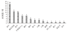
Figure 1
Intracranial distribution of abnormal MRI signals in 34 patients with acute lymphoblastic leukemia with chemotherapy-related cerebral lesion in encephalopathy group"

| [1] |
Jeha S, Pei D, Choi J, et al. Improved CNS control of childhood acute lymphoblastic leukemia without cranial irradiation: St Jude total therapy study 16[J]. J Clin Oncol, 2019, 37(35): 3377-3391.
doi: 10.1200/JCO.19.01692 pmid: 31657981 |
| [2] | Taillibert S, Le Rhun E, Chamberlain MC. Chemotherapy-related neurotoxicity[J]. Curr Neurol Neurosci Rep, 2016, 16(9): 81. |
| [3] |
Gibson EM, Nagaraja S, Ocampo A, et al. Methotrexate chemotherapy induces persistent tri-glial dysregulation that underlies chemotherapy-related cognitive impairment[J]. Cell, 2019, 176(1-2): 43-55.
doi: S0092-8674(18)31405-3 pmid: 30528430 |
| [4] |
Lenfant C, Greiner N, Duprez T. Cytarabine-induced encephalitis[J]. J Belg Soc Radiol, 2021, 105(1): 46.
doi: 10.5334/jbsr.2492 pmid: 34611580 |
| [5] |
Newey CR, Chandrasekaran PN, Mohebbi MR. Posterior reversible encephalopathy syndrome after high-dose cytarabine in acute myelogenous leukemia[J]. Neurol India, 2017, 65(1): 220.
doi: 10.4103/0028-3886.198171 pmid: 28084289 |
| [6] |
Abaji R, Krajinovic M. Pharmacogenetics of asparaginase in acute lymphoblastic leukemia[J]. Cancer Drug Resist, 2019, 2(2): 242-255.
doi: 10.20517/cdr.2018.24 pmid: 35582721 |
| [7] |
Magge RS, DeAngelis LM. The double-edged sword: neurotoxicity of chemotherapy[J]. Blood Rev, 2015, 29(2): 93-100.
doi: 10.1016/j.blre.2014.09.012 pmid: 25445718 |
| [8] | 吴晔. 儿童脑白质病变的识别及诊断[J]. 北京医学, 2018, 40(7): 614-615. |
| Wu Y. Identification and diagnosis of white matter lesions in children[J]. Beijing Yikue, 2018, 40(7): 614-615. | |
| [9] | 中华医学会儿科学分会血液学组, 《中华儿科杂志》编辑委员会. 儿童急性淋巴细胞白血病诊疗建议(第四次修订)[J]. 中华儿科杂志, 2014, 52(9): 641-644. |
| Hematology Group of pediatric Society of Chinese Medical Association, Editorial Board of Chinese Journal of Pediatrics. Recommendations for diagnosis and treatment of childhood acute lymphoblastic leukemia (the fourth revision)[J]. Zhonghua Erke Zazhi, 2014, 52(9): 641-644. | |
| [10] |
Conklin HM, Krull KR, Reddick WE, et al. Cognitive outcomes following contemporary treatment without cranial irradiation for childhood acute lymphoblastic leukemia[J]. J Natl Cancer Inst, 2012, 104(18): 1386-1395.
pmid: 22927505 |
| [11] | Cao SC, Legerstee JS, van Bellinghen M, et al. Effect of chemotherapy (with and without radiotherapy) on the intelligence of children and adolescents treated for acute lymphoblastic leukemia; a meta-analysis[J]. Psychooncology, 2023, 32(4): 492-505. |
| [12] | Jacola LM, Conklin HM, Krull KR, et al. The impact of intensified CNS-directed therapy on neurocognitive outcomes in survivors of childhood acute lymphoblastic leukemia treated without cranial irradiation[J]. J Clin Oncol, 2022, 40(36): 4218-4227. |
| [13] |
Krull KR, Cheung YT, Liu W, et al. Chemotherapy pharmacodynamics and neuroimaging and neurocognitive outcomes in long-term survivors of childhood acute lymphoblastic leukemia[J]. J Clin Oncol, 2016, 34(22): 2644-2653.
doi: 10.1200/JCO.2015.65.4574 pmid: 27269941 |
| [14] | Śliwa-Tytko P, Kaczmarska A, Lejman M, et al. Neurotoxicity associated with treatment of acute lymphoblastic leukemia chemotherapy and immunotherapy[J]. Int J Mol Sci, 2022, 23(10): 5515. |
| [15] | Sainz de la Maza Cantero S, Jiménez Martín A, de Felipe Mimbrera A, et al. Cerebellar toxicity due to cytarabine: a series of 4 cases[J]. Neurologia, 2016, 31(7): 491-492. |
| [16] |
Drachtman RA, Cole PD, Golden CB, et al. Dex-tromethorphan is effective in the treatment of subacute methotrexate neurotoxicity[J]. Pediatr Hematol Oncol, 2002, 19(5): 319-327.
pmid: 12078863 |
| [17] |
Smith GA, Damon LE, Rugo HS, et al. High-dose cytarabine dose modification reduces the incidence of neurotoxicity in patients with renal insufficiency[J]. J Clin Oncol, 1997, 15(2): 833-839.
pmid: 9053511 |
| [18] |
Partap S, Russo S, Esfahani B, et al. A review of chronic leukoencephalopathy among survivors of childhood cancer[J]. Pediatr Neurol, 2019, 101: 2-10.
doi: S0887-8994(18)31330-4 pmid: 31047756 |
| [19] |
Caruso V, Iacoviello L, Di Castelnuovo A, et al. Thrombotic complications in childhood acute lymphoblastic leukemia: a meta-analysis of 17 prospective studies comprising 1752 pediatric patients[J]. Blood, 2006, 108(7): 2216-2222.
doi: 10.1182/blood-2006-04-015511 pmid: 16804111 |
| [20] |
Kieslich M, Porto L, Lanfermann H, et al. Cerebrovascular complications of L-asparaginase in the therapy of acute lymphoblastic leukemia[J]. J Pediatr Hematol Oncol, 2003, 25(6): 484-487.
pmid: 12794528 |
| [21] |
Goyal G, Bhatt VR. L-asparaginase and venous thromboembolism in acute lymphocytic leukemia[J]. Future Oncol, 2015, 11(17): 2459-2470.
doi: 10.2217/fon.15.114 pmid: 26274336 |
| [22] | Alessi I, Caroleo AM, de Palma L, et al. Short and long-term toxicity in pediatric cancer treatment: central nervous system damage[J]. Cancers (Basel), 2022, 14(6): 1540. |
| [23] | Kim KH, Park M, Park EY, et al. Disseminating necrotizing leukoencephalopathy associated with intra-CSF methotrexate chemotherapy: a retrospective observational study[J]. Neurology, 2024, 102(5): e209167. |
| [1] | HE Ying, LIU Zhiyong, YANG Hansong, CAI Yali, XU Jinglin, CHEN Dongmei. Clinical analysis of 153 neonatal enterovirus infections and antibiotic management improvement study [J]. Journal of Clinical Pediatrics, 2025, 43(2): 128-134. |
| [2] | CHU Sijia, TANG Jihong. Research progress of central nervous system injury associated with pediatric acute lymphoblastic leukemia and its treatment [J]. Journal of Clinical Pediatrics, 2024, 42(9): 811-816. |
| [3] | LU Jun. The clinical significance and management strategies of B cell aplasia following CD19 CAR-T cell therapy in pediatric patients [J]. Journal of Clinical Pediatrics, 2024, 42(7): 578-582. |
| [4] | WANG Yu, XUE Yujuan, ZUO Yingxi, JIA Yueping, LU Aidong, ZENG Huimin, ZHANG Leping. Efficacy and safety of CD19 targeted CAR-T cells in the treatment of refractory/relapsed B-cell acute lymphoblastic leukemia in children and adolescents [J]. Journal of Clinical Pediatrics, 2024, 42(7): 583-588. |
| [5] | WANG Yuejia, XIA Lei, CHENG Huiqing, LYU Rongji, FAN Kun, XU Falin. Clinical characteristics of extremely premature infants conceived by assisted reproductive technology [J]. Journal of Clinical Pediatrics, 2024, 42(7): 600-605. |
| [6] | ZHONG Xiaomei, REN Hong, ZHANG Jian, LI Biru, QIAN Juan, NING Botao, GAO Yijin, WANG Ying. Comparison of clinical features between hemophagocytic lymphohistiocytosis and sepsis [J]. Journal of Clinical Pediatrics, 2024, 42(6): 547-552. |
| [7] | CHEN Shicai, DUAN Lifen, SUN Ying, SHAO Juwei, LI Qiong, LUO Mingying, REN Junjun, ZHANG Yunqian. Correlation between cognitive function and electroclinical characteristics of benign childhood epilepsy with centrotemporal spikes [J]. Journal of Clinical Pediatrics, 2024, 42(3): 211-217. |
| [8] | HUANG Shihao, YUAN Xiaojun. Analysis on clinical characteristics and treatment for Kasabach-Merritt phenomenon in 36 children with hemangioma [J]. Journal of Clinical Pediatrics, 2024, 42(11): 917-921. |
| [9] | YANG Liu, SU Meng, ZHANG Jing, AN Kang, CAI Jiaoyang, QIAN Juan, TANG Yanjing, LI Benshang. Clinical analysis of CD19/CD22 CAR-T cell therapy for MLL gene rearrangement-positive refractory/relapsed childhood acute B-lineage lymphoblastic leukemia [J]. Journal of Clinical Pediatrics, 2024, 42(10): 888-894. |
| [10] | HOU Ruolin, WU Jing, LI Ling. Pediatric autoimmune encephalitis with brain MRI showing meningeal thickening and enhancement [J]. Journal of Clinical Pediatrics, 2023, 41(9): 674-679. |
| [11] | WU Xiaoling, LYU Tiewei. Clinical analysis of idiopathic left ventricular tachycardia in children [J]. Journal of Clinical Pediatrics, 2023, 41(8): 599-603. |
| [12] | YU Liting, SHEN Xingwei, WANG Zhuo, ZHANG Shunguo, GAO Yijin. Efficacy and safety of fosaprepitant in the prevention of highly emetogenic chemotherapy-related nausea and vomiting in pediatric patients with cancer [J]. Journal of Clinical Pediatrics, 2023, 41(8): 604-609. |
| [13] | XUE Yujuan, LU Aidong, WANG Yu, JIA Yueping, ZUO Yingxi, ZHANG Leping. Clinical analysis of treatment failure in children with acute lymphoblastic leukemia [J]. Journal of Clinical Pediatrics, 2023, 41(3): 204-209. |
| [14] | HUA Minmin, XIA Lei, HUO Wanying, ZHANG Yanhua, XU Falin. Analysis of clinical and prognostic factors of neonatal congenital chylothorax [J]. Journal of Clinical Pediatrics, 2023, 41(1): 25-29. |
| [15] | SHI Yan, YANG Meng, HUANG Yitian, XU Junjie, WEN Sheng, HE Dawei, WEI Guanghui, HUA Yi. Therapeutic effect of chemotherapy on lung metastases of nephroblastoma [J]. Journal of Clinical Pediatrics, 2022, 40(9): 696-700. |
|
||