临床儿科杂志 ›› 2022, Vol. 40 ›› Issue (4): 258-262.doi: 10.12372/jcp.2022.21e0199

• 呼吸系统疾病专栏 • 上一篇    下一篇

大叶性肺炎形成支气管黏液栓高危因素分析

苏艳艳, 汤昱(), 王艳琼, 徐沙沙, 董利利   

  1. 郑州大学附属儿童医院 河南省儿童医院 郑州儿童医院呼吸科(河南郑州 450000)
  • 收稿日期:2021-02-05 出版日期:2022-04-15 发布日期:2022-04-07
  • 通讯作者: 汤昱 E-mail:tangyu010312@sina.com

Analysis of high-risk factors of bronchial mucus plugs formation in children with lobar pneumonia

SU Yanyan, TANG Yu(), WANG Yanqiong, XU Shasha, DONG Lili   

  1. Department of Respiratory Medicine, Henan Children's Hospital, Zhengzhou Children's Hospital, Zhengzhou 450000, Henan, China
  • Received:2021-02-05 Online:2022-04-15 Published:2022-04-07
  • Contact: TANG Yu E-mail:tangyu010312@sina.com

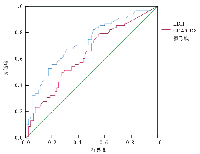
摘要: 目的 探讨大叶性肺炎形成支气管黏液栓的临床特征和高危因素。方法 选取2018年1月至2020年3月收治的大叶性肺炎患儿临床资料。根据纤维支气管镜下情况分为黏液栓组和非黏液栓组,比较两组患儿临床特征和实验室检查结果。结果 共收治大叶性肺炎患儿935例。根据纤维支气管镜检查结果纳入黏液栓组135例,男73例、女62例,中位年龄5.0(3.1~7.0)岁;简单随机法抽取非黏液栓组135例,男75例、女60例,中位年龄5.8(4.0~7.0)岁。与非黏液栓组相比,黏液栓组的热程较长,中性粒细胞百分比、C-反应蛋白(CRP)、降钙素原(PCT)、乳酸脱氢酶(LDH)、D-二聚体、CD4/CD8水平较高,血小板计数较低,差异均有统计学意义(P<0.05)。两组病原体均以肺炎支原体为主,黏液栓组肺炎支原体感染比例为92.6%,肺炎支原体合并细菌感染比例为20.7%,肺炎支原体合并病毒感染比例为51.1%,肺炎支原体合并EB病毒感染比例为14.1%,高于非黏液栓组的77.0%、11.9%、20.0%、0.7%,差异有统计学意义(χ2=3.91~28.51,P均<0.05)。黏液栓组感染部位为肺下叶和左肺下叶的比例高于非黏液栓组,胸腔积液、肝功能损伤以及后遗症发生率均高于非黏液栓组,差异有统计学意义(P<0.05)。二分类logistic回归分析结果提示,LDH和CD4/CD8是大叶性肺炎形成黏液栓的独立危险因素(P<0.05)。结论 大叶性肺炎患儿支气管黏液栓的形成与LDH、CD4/CD8有关。

关键词: 大叶性肺炎, 支气管黏液栓, 临床特征, 高危因素, 儿童

Abstract: Objective To investigate the clinical characteristics and high-risk factors of bronchial mucus plugs formation in children with lobar pneumonia.Methods Children with lobar pneumonia treated from January 2018 to March 2020 were selected as the study subjects. According to the results of fiberoptic bronchoscopy, the children were divided into the mucous plugs group and the non-mucous plugs group, and the clinical characteristics and laboratory examination results of the two groups were compared.Results A total of 935 children with lobar pneumonia were treated. According to fiberoptic bronchoscopy, 135 patients (73 boys and 62 girls) were included in the mucous plugs group, and the median age was 5.0 (3.1-7.0) years. There were 135 children (75 boys and 60 girls) in the non-mucous plugs group, and the median age was 5.8 (4.0-7.0) years. Compared with the non-mucous plugs group, the mucous plugs group had a longer fever duration, a higher percentage of neutrophil, higher levels of c-reactive protein (CRP), procalcitonin (PCT), lactate dehydrogenase (LDH), D-2 dimer and CD4/CD8, and a lower platelet count, with statistical significance (P<0.05). Mycoplasma pneumoniae was the main pathogen in both groups. The proportions of Mycoplasma pneumoniae infection, Mycoplasma pneumoniae combined with bacterial infection, Mycoplasma pneumoniae combined with virus infection and Mycoplasma pneumoniae combined with Epstein-Barr virus infection in the mucous plugs group were 92.6%, 20.7%, 51.1% and 14.1%, respectively, which were higher than 77.0%, 11.9%, 20.0% and 0.7% in the non-mucous plugs group, and the differences were statistically significant (χ2=3.91-28.51, P<0.05). The proportion of infection sites in the lower lobe of lung and left lower lobe of lung in the mucus plugs group was higher than that in the non-mucus plugs group, and the incidence of pleural effusion, liver function injury and sequelae was higher than that in the non-mucus plugs group, and the differences were statistically significant (P<0.05). The results of binary logistic regression analysis suggested that LDH and CD4/CD8 were independent risk factors for the formation of mucous plugs in lobar pneumonia (P<0.05).Conclusions The formation of bronchial mucus plugs in children with lobar pneumonia is related to LDH and CD4/CD8.

Key words: lobar pneumonia, bronchial mucus plug, clinical feature, risk factor, child