临床儿科杂志 ›› 2022, Vol. 40 ›› Issue (9): 685-689.doi: 10.12372/jcp.2022.21e1338

• 新生儿疾病专栏 • 上一篇    下一篇

新生儿脑白质静脉梗死8例临床分析

林雅茵(), 郑直1, 赵锋2   

  1. 1.新生儿科,厦门大学附属妇女儿童医院(厦门市妇幼保健院)(福建厦门 361000)
    2.放射科 厦门大学附属妇女儿童医院(厦门市妇幼保健院)(福建厦门 361000)
  • 收稿日期:2021-09-16 出版日期:2022-09-15 发布日期:2022-08-26
  • 通讯作者: 林雅茵 E-mail:linyy0806@163.com

Clinical analysis of 8 neonates with cerebral white matter venous infarction

LIN Yayin(), ZHENG Zhi1, ZHAO Feng2   

  1. 1. Department of Neonatology, School of Medicine, Xiamen University, Xiamen 361000, Fujian, China
    2. Department of Radiology, Women and Children’s Hospital, School of Medicine, Xiamen University, Xiamen 361000, Fujian, China
  • Received:2021-09-16 Online:2022-09-15 Published:2022-08-26
  • Contact: LIN Yayin E-mail:linyy0806@163.com

摘要:

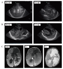
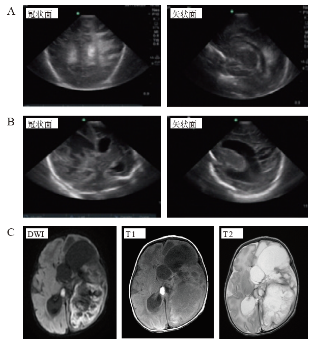
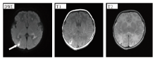
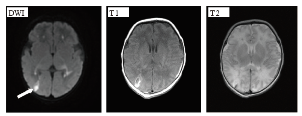
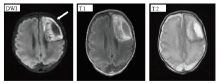
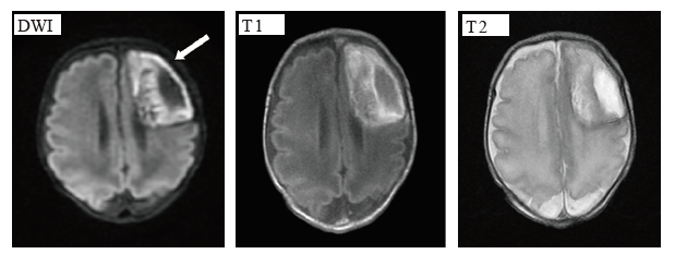
目的 探讨新生儿深部脑白质静脉梗死的发病因素、临床表现及影像学特征。方法 回顾分析经影像学确诊的8例新生儿深部脑白质静脉梗死患者的临床资料,分析其发病因素、临床表现及影像学特征。结果 8例患儿中6例为早产儿、2例为足月儿,平均胎龄(32.2±3.9)周,平均出生体重(1 932±794)g,发病日龄1~30 d。5例行胎盘病理检查,4例提示绒毛膜羊膜炎、1例绒毛间隙血栓,4例合并化脓性脑膜炎,3例存在围生期缺氧史,1例为宫内风疹病毒感染伴红细胞增多症。4例患儿有发热,3例短暂惊厥发作,2例无症状。1例患儿行颅脑梗死部位穿刺,抽出血水样液体60 mL,细菌培养阴性。在发病第2~54天行头颅MRI检查,显示脑白质静脉梗死部位多发于额顶叶白质(6/8),病变周围都有增强信号,脑梗死部位可伴液化、空洞,与脑脓肿极易混淆。结论 新生儿深部脑白质静脉梗死的诱发因素主要有绒毛膜羊膜炎、化脓性脑膜炎及围生期缺氧,临床表现易被忽略,MRI表现易与脑脓肿混淆,应注意鉴别。

关键词: 新生儿, 脑白质静脉梗死, 脑脓肿

Abstract:

Objective To investigate the risk factors, clinical manifestations and imaging characteristics of cerebral deep white matter venous infarction in neonates. Methods The clinical data of 8 neonates with cerebral deep white matter venous infarction confirmed by cranial imaging during January 1, 2017 to December 31, 2019 were retrospectively analyzed, and the etiological factors, clinical manifestations and imaging characteristics of the neonates were analyzed and summarized. Results Among the 8 neonates with deep white matter venous infarction, 6 were premature and 2 were full term. The mean gestational age was (32.2±3.9) weeks and the mean birth weight was (1932.0±794.0) g. The age of onset ranged from 1 day to 30 days. Five neonates underwent pathological examination of placenta, including 4 cases of chorioamnionitis and 1 case of intervillous thrombus. Four cases were complicated with purulent meningitis, 3 cases had a history of perinatal hypoxia, and 1 case had intrauterine rubella virus infection with polycythemia. Among the 8 neonates, 4 had fever, 3 had transient seizures, and 2 were asymptomatic. One patient underwent puncture at the site of craniocerebral infarction, and 60mL of bleeding like fluid was aspirated, and bacterial culture was negative. Cranial MRI was performed between 2 and 54 days after onset. MRI showed that white matter venous infarction mostly occurred in the frontal parietal white matter (6/8), with enhanced signals surrounding the lesions. Cerebral infarction site could be accompanied by liquefaction and cavity, which is easily confused with cerebral abscess. Conclusions Chorioamnionitis, purulent meningitis and perinatal hypoxia are the main inducing factors of neonatal deep white matter venous infarction. The clinical manifestations of cerebral venous infarction are easy to be ignored. Their MRI findings are often confused with cerebral abscesses, and attention should be paid tothe identification.

Key words: newborn, cerebral white matter venous infarction, cerebral abscesses