临床儿科杂志 ›› 2026, Vol. 44 ›› Issue (8): 698-704.doi: 10.12372/jcp.2026.25e1347

• 临床研究 • 上一篇    下一篇

儿童失张力发作的临床与电生理特征分析

张晓, 方红军, 旷小军, 王丽娟, 吴照, 蒋莎, 李佩, 吴丽文()   

  1. 中南大学湘雅医学院附属儿童医院(湖南省儿童医院)神经内科(湖南长沙 410007)
  • 收稿日期:2025-11-03 修回日期:2026-02-28 录用日期:2026-03-18 出版日期:2026-08-15 发布日期:2026-08-03
  • 通讯作者: 吴丽文 E-mail:wuliwen2020@sina.com
  • 作者简介:第一联系人:

    张晓、方红军收集、分析数据并撰写论文初稿,王丽娟、吴照、蒋莎、李佩采集及分析数据、统计数据、审阅文章,旷小军、吴丽文、方红军负责选题、研究设计及论文修改。

  • 基金资助:
    湖南省中医药重点研究课题(A2024042);湖南省杰出青年科学基金项目(2024JJ2035)

Analysis of the clinical and electrophysiological characteristics of atonic seizures in children

ZHANG Xiao, FANG Hongjun, KUANG Xiaojun, WANG Lijuan, WU Zhao, JIANG Sha, LI Pei, WU Liwen()   

  1. Department of Neurology, Affiliated Children's Hospital of Xiangya Medical College, Central South University/Hunan Children's Hospital, Changsha 410007, Hunan, China
  • Received:2025-11-03 Revised:2026-02-28 Accepted:2026-03-18 Published:2026-08-15 Online:2026-08-03
  • Contact: WU Liwen E-mail:wuliwen2020@sina.com

摘要:

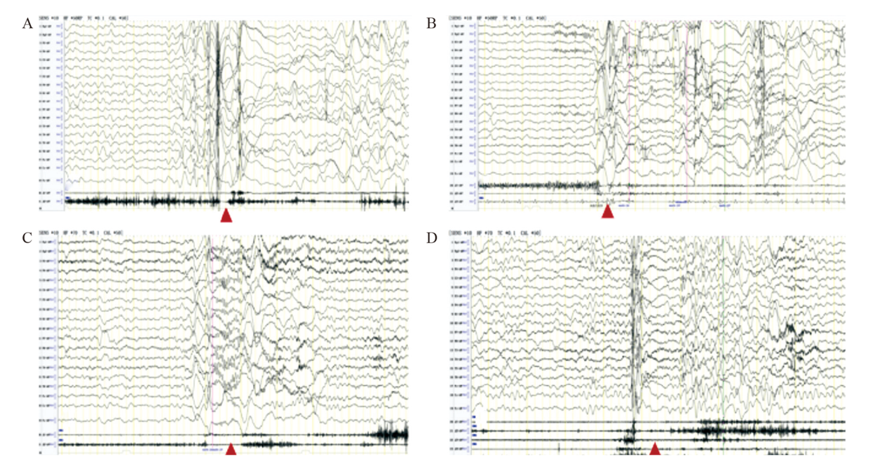
目的 失张力发作没有先兆,患儿可能会反复跌倒,严重影响其安全和生活质量。失张力发作的准确诊断和管理较为困难,发作期脑电及肌电特征是鉴别诊断的金标准。本研究总结了存在失张力发作的癫痫患儿的临床、脑电图和肌电图特征、治疗及预后,以期为临床医师正确诊治此类疾病提供参考。方法 回顾性分析神经内科2015年8月至2023年12月收治的失张力发作患儿的临床资料,分析总结患儿的临床特点、病因、治疗和随访情况。结果 共47例患儿存在失张力发作,男32例、女15例,中位起病年龄为2.17(1.17~2.92)岁,其中结构性病因7例、遗传性病因10例、遗传性结构病因1例、病因未明29例。失张力发作类型包括全面性失张力发作(33例)、肌阵挛-失张力发作(5例)、痉挛-失张力发作(2例)、不典型失神伴失张力发作(2例)、失张力发作和肌阵挛-失张力发作均存在者(4例)、局灶性失张力发作(1例)。患儿常伴有其他发作类型,以肌阵挛发作为主(15/47),其次为不典型失神发作(6/47)、痉挛发作(5/47)等。47例患儿中13例明确癫痫综合征类型,其中癫痫伴肌阵挛失张力发作(EMAS)6例、婴儿癫痫性痉挛综合征(IESS)5例、Lennox-Gastaut综合征(LGS)1例、Dravet综合征(DS)1例。47例患儿中除5例发育正常外,其余42例均存在一定程度智力障碍。失张力发作期脑电图表现为棘慢波20例、慢波13例、多棘慢波8例、低波幅快节律2例、慢波复合低波幅快波2例、慢波后紧随低波幅快节律1例,脑电图无改变1例(仅表现肌电静息)。发作期肌电图表现为肌电抑制43例,有4例肌电抑制不明显。47例患儿中,27例控制至少6个月无发作,14例发作次数减少50%,6例抗癫痫治疗后无效。结论 失张力发作男性多于女性,可合并多种发作形式,见于多种癫痫综合征中,病因以遗传和结构性病因为主,大多伴有智力障碍。发作期脑电图主要表现为(多)棘慢波、慢波、低波幅快节律、慢波复合低波幅快波、慢波后紧随低波幅快节律以及脑电图无改变,同步肌电图监测对发现失张力发作很重要。

关键词: 失张力发作, 癫痫, 脑电图, 癫痫综合征, 儿童

Abstract:

Objective Atonic seizures occur without warning, and children with this condition may repeatedly fall, seriously affecting their safety and quality of life. Accurate diagnosis and management of atonic seizures are rather difficult, and the electroencephalography (EEG) and electromyography (EMG) features during the seizure period are the gold standard for differential diagnosis. This study summarizes the clinical, EEG, and EMG characteristics, treatment, and prognosis of children with epilepsy who have atonic seizures, with the aim of providing a reference for clinicians to correctly diagnose and treat this disease. Methods A retrospective analysis was conducted on the clinical data of children with atonic seizures admitted to the neurology department from August 2015 to December 2023. The clinical characteristics, etiology, treatment and follow-up of the children were analyzed and summarized. Results A total of 47 children had atonic seizures, including 32 boys and 15 girls. The median age of onset was 2.17 (1.17-2.92) years. Among them, 7 children had structural causes, 10 had hereditary causes, 1 had hereditary structural cause, and the causes were unknown in 29 children. The types of atonic seizures included generalized atonic seizures (33 cases), myoclonic-atonic seizures (5 cases), spasm-atonic seizures (2 cases), atypical absence seizures with atonic seizures (2 cases), both atonic seizures and myoclonic-atonic seizures (4 cases), and focal atonic seizures (1 case). The patients often have other types of seizures, with myoclonic seizures being the predominant type (15/47, 31.9%), followed by atypical absence seizures (6/47), and spasms (5/47), etc. Among the 47 children, the type of epileptic syndromes was clearly identified in 13 cases. Among them, 6 cases had epilepsy with myoclonic atonic seizures (EMAS), 5 had infantile epileptic spasms syndrome (IESS), 1 had Lennox-Gastaut syndrome (LGS), and 1 had Dravet syndrome (DS). Among the 47 patients, except for 5 patients with normal development, the remaining 42 all have some degree of intellectual disability. During atonic seizures, EEG manifestations included spike-slow waves in 20 cases, slow waves in 13 cases, multispike-slow waves in 8 cases, low-amplitude fast rhythms in 2 cases, slow waves composite fast waves in 2 cases, and slow waves followed by low-amplitude fast rhythms in 1 case. No EEG changes were observed in 1 case (electromyographic rest only). During seizures, EMG showed electromyographic suppression in 43 cases, while electromyographic suppression was inconspicuous in 4 cases. Among the 47 patients, 27 patients were controlled for at least 6 months without seizures, 14 patients had a 50% reduction in seizure frequency, and 6 patients were ineffective after anti-seizure medication. Conclusions Atonic seizures are more common in males than females and can be combined with multiple seizure forms, which are common in various epileptic syndromes. The etiology is mainly genetic and structural diseases, most of which are accompanied by intellectual disability. The main manifestations of ictal EEG are (multiple) spike slow waves, slow waves, low amplitude fast rhythms, slow wave composite fast waves, and slow waves followed closely by fast rhythms, and unchanged in EEG. Synchronous EMG monitoring is important for detecting atonic seizures.

Key words: atonic seizures, epilepsy, electroencephalography, epileptic syndromes, child

中图分类号: 

  • R72