临床儿科杂志 ›› 2024, Vol. 42 ›› Issue (12): 1047-1050.doi: 10.12372/jcp.2024.24e0285

• 临床报道 • 上一篇    下一篇

一例弥漫性肺淋巴管瘤病支气管镜下表现及分析

曾祥妮1, 吴爱民1(), 李岚1, 涂洪强2, 周俊霖3, 樊金星4   

  1. 1.江西省儿童医院 呼吸内科,(江西南昌 330038)
    2.江西省儿童医院 小儿心脏病治疗中心,(江西南昌 330038)
    3.江西省儿童医院 医学放射科,(江西南昌 330038)
    4.江西省儿童医院 医学病理科(江西南昌 330038)
  • 收稿日期:2024-04-02 出版日期:2024-12-15 发布日期:2024-12-02
  • 通讯作者: 吴爱民 电子信箱: wamzjy@163.com
  • 基金资助:
    江西省卫生健康委科技计划(202211205)

A case report of a rare case of diffuse pulmonary lymphangiomatosis with bronchoscopic presentation

ZENG Xiangni1, WU Aimin1(), LI Lan1, TU Hongqiang2, ZHOU Junlin3, FAN Jinxing4   

  1. 1. Department of Respiratory Medicine, Jiangxi, China
    2. Pediatric Heart treatment Center, Jiangxi, China
    3. Medical Radiology Department, Jiangxi, China
    4. Medical Pathology Department, Jiangxi Children's Hospital, Nanchang 330038, Jiangxi, China
  • Received:2024-04-02 Published:2024-12-15 Online:2024-12-02

摘要:

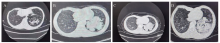
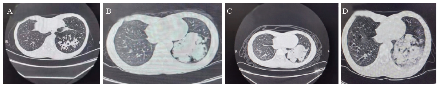
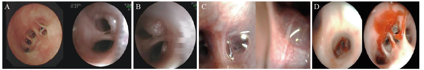
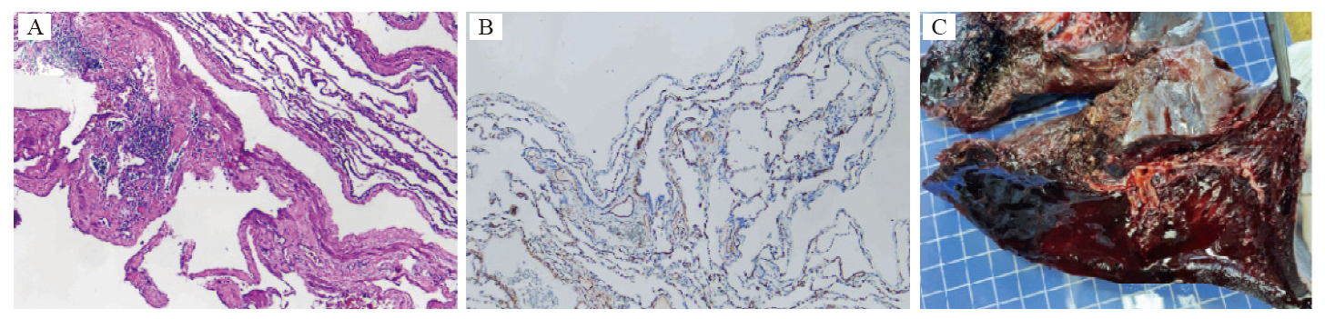
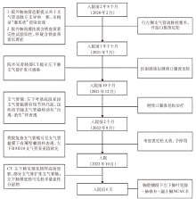
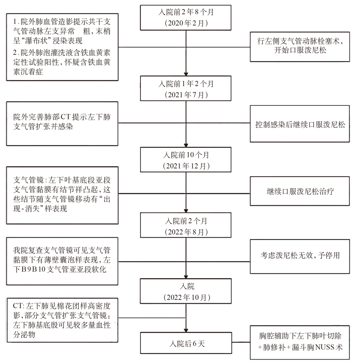
回顾性分析1例来弥漫性肺淋巴管瘤病(DPL)的支气管镜下表现,为DPL早期诊断提供参考。患儿,男,因“间断咯血2年8个月”来我院就诊,既往行左侧支气管动脉栓塞术后咯血无改善,多次气管镜下灌洗液含铁血黄素定性试验阳性,予口服激素治疗2年余仍反复咯血,最终切除患肺行病理检查提示DPL。本文总结患儿支气管镜下表现,发现早期该患儿镜下可见结节样凸起,这些结节随支气管镜移动有“出现-消失”样表现,类似活瓣,间隔9个月后复查支气管镜可见相同部位支气管黏膜下有薄壁囊泡样表现,同时支气管亚段有外压表现。说明支气管镜检查可为DPL早期发现提供参考。

关键词: 弥漫性肺淋巴管瘤病, 支气管镜, 咯血, 青春期

Abstract:

This study retrospectively analyzed the bronchoscopic manifestations in a pediatric case of diffuse pulmonary lymphangiomatosis (DPL), aiming to provide references for its early diagnosis. The child came to our hospital in August presented with “intermittent hemoptysis for 2 years and 8 months”, Despite previous left bronchial artery embolization, the hemoptysis persisted., and the qualitative test of ferritin was positive in several times of tracheoscopic lavage. After more than 2 years of oral corticosteroid therapy, the patient continued to experience recurrent hemoptysis.Ultimately, lung resection and pathological examination confirmed the diagnosis of DPL.This paper summarizes the bronchoscopic findings, noting that in the early stages, nodular protrusions visible under the bronchoscope exhibited an “appear-disappear” phenomenon, resembling a valve. Nine months later, a follow-up bronchoscopy revealed thin-walled vesicular manifestations under the bronchial mucosa at the same site, along with external compression of bronchial subsegments. These findings suggest that bronchoscopy can provide valuable insights for the early detection of DPL.

Key words: diffuse pulmonary lymphangiomatosis, bronchoscopy, haemoptysis, adolescent